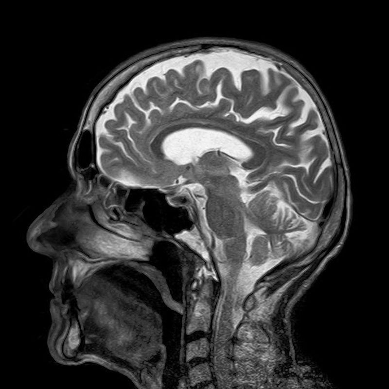
From Infinity to the Brain

We have watched the science of medical imaging grow so much over the last several decades that perhaps we should not be surprised at another potentially huge leap in this technology. But, when you start talking about using space born technology here on earth to help cure brain tumors, that is exciting.
Medical imaging has grown to include a wide array of technologies from sonograms (ultrasound pictures) to PET scans (positron emission tomography), and MRI (magnetic resonance imaging). The hope is that Ultraviolet Light Imaging can look deeper into tissue and help surgeons make better decisions about what is and is not tumor tissue.
Key Takeaways
- Researchers at Cedars-Sinai are repurposing ultraviolet telescope camera technology for brain surgery.
- The technology is used to differentiate between tumor cells and healthy brain tissue in real-time during surgery.
- Ultraviolet light emitted by specific chemicals present in tumor cells can be captured by the camera.
- This innovative approach aims to improve the precision of brain tumor removal, reducing the chances of leaving behind active tumor cells.
- The trial is still in progress, with the hope that data collected will refine the imaging process for future applications in surgery.
Planet-Exploring Technology in the Operating Room
A clinical trial at Cedars-Sinai Medical Center and the Maxine Dunitz Neurosurgical Institute are using camera technology from the ultraviolet telescopes initially designed to study suns, planets and galaxies, to help surgeons better define brain tumors. They are beginning by focusing on glioma tumors. Glial cells from which glioma tumors grow are five times more prevalent in the brain than even neurons. Unlike neurons, glial cells have the ability to divide and multiply.
One of chief concerns among neurosurgeons when operating in the brain is to be able to identify tumor cells from healthy cells. This allows them to take as little tissue as possible and still get as much of the tumor as is practical. If they leave some tumor cells behind these cells can continue to multiply and recreate the tumor. These kinds of tumors often multiply as tentacles growing through healthy tissue, not just lumps.
According to Dr. Ray Chu, one of the co-leaders of the clinical trial, “The ultraviolet imaging technique may provide a ‘metabolic map’ of tumors that could help us differentiate them from normal surrounding brain tissue, providing useful, real-time, intraoperative information.” In the trial the ultraviolet camera is placed as close to the operating field as possible to perform the imaging. In the trial, decisions are not being made from the data the imaging provides but instead is looked at post surgery along with all the other medical imaging and laboratory data. The team can then begin to assess the value of the information and how to set up the imaging protocols for the future.
What They Hope to See
Ultraviolet light in nature helps some insects to find pollen, for instance, on some flowers. Their eyesight extends into the ultraviolet and can provide them information about where good food is, like the following image.
Tumor cells are much more active and have certain chemicals. One example, a specific chemical (nicotinamide adenine dinucleotide hydrogenase or NADH) accumulates in brain tumor cells but not in healthy cells. NADH emits ultraviolet light that can be differentiated by this ultraviolet imaging process. The clinical trial is one way to start collecting data so the imaging camera can be fine-tuned to see the edges of the tumors.
Conclusion
Camera technology from a space born telescope looking into infinity can very likely be modified to help neurosurgeons here on earth using what nature provides us in the form of ultraviolet light. We know that ultraviolet light affects many living things on earth including human tissue. Might this technology begin to help us define the very building blocks of memory so we could help stroke victims and the recovery of other memory loss? We don’t know yet, but the hope is that technology is now being refined.
Reference Article: Galaxy-Exploring Camera to Be Used in the Operating Room.
Would you like to receive similar articles by email?


